11.3 Periosteal chondroma
– Jeremy C. Collette, MD, PhD
__________________________________________________________________
11.3.1 Background
Periosteal chondroma, also known as juxtacortical chondroma, is a rare benign surface lesion of bone with mature hyaline cartilage differentiation. It is generally more cellular than an enchondroma.
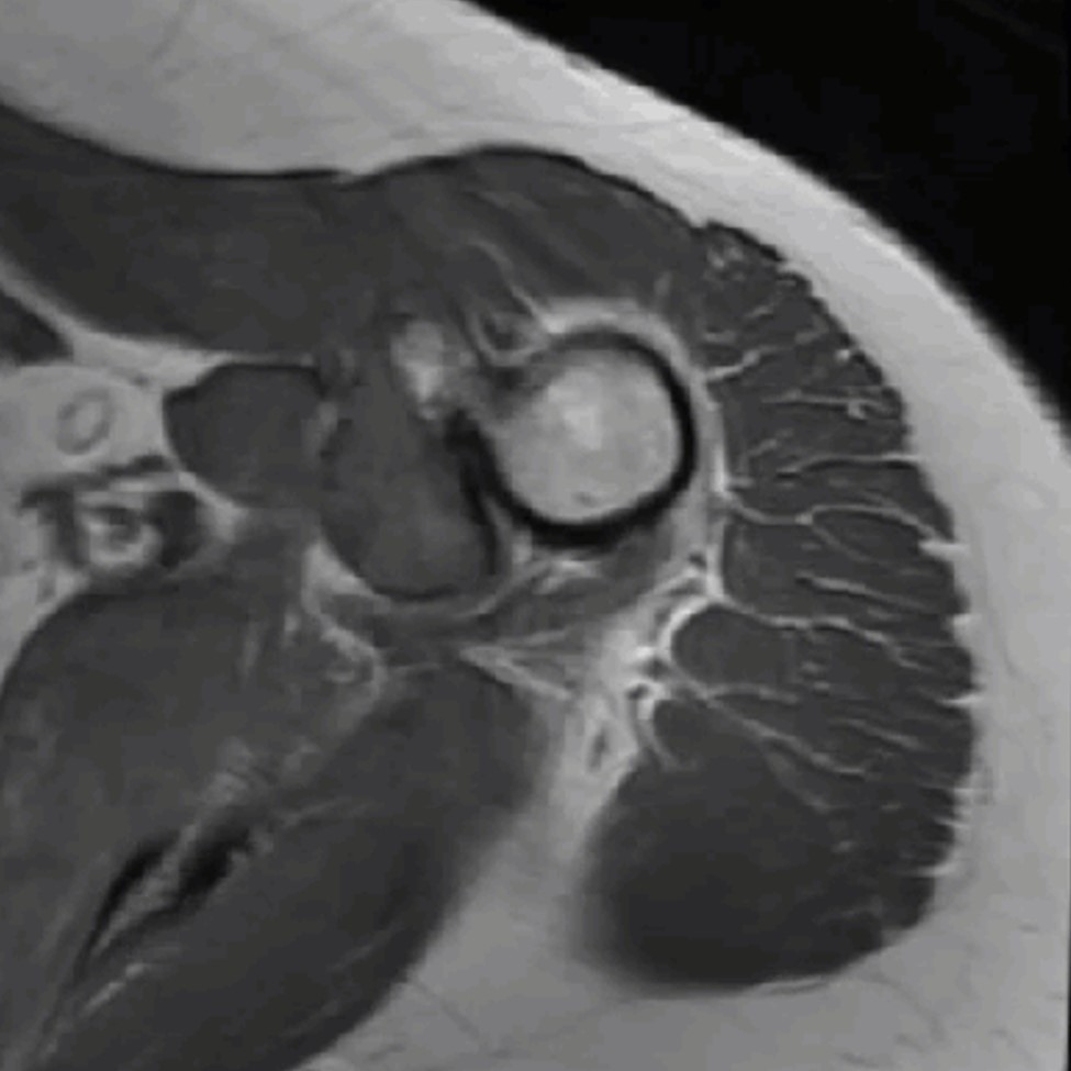
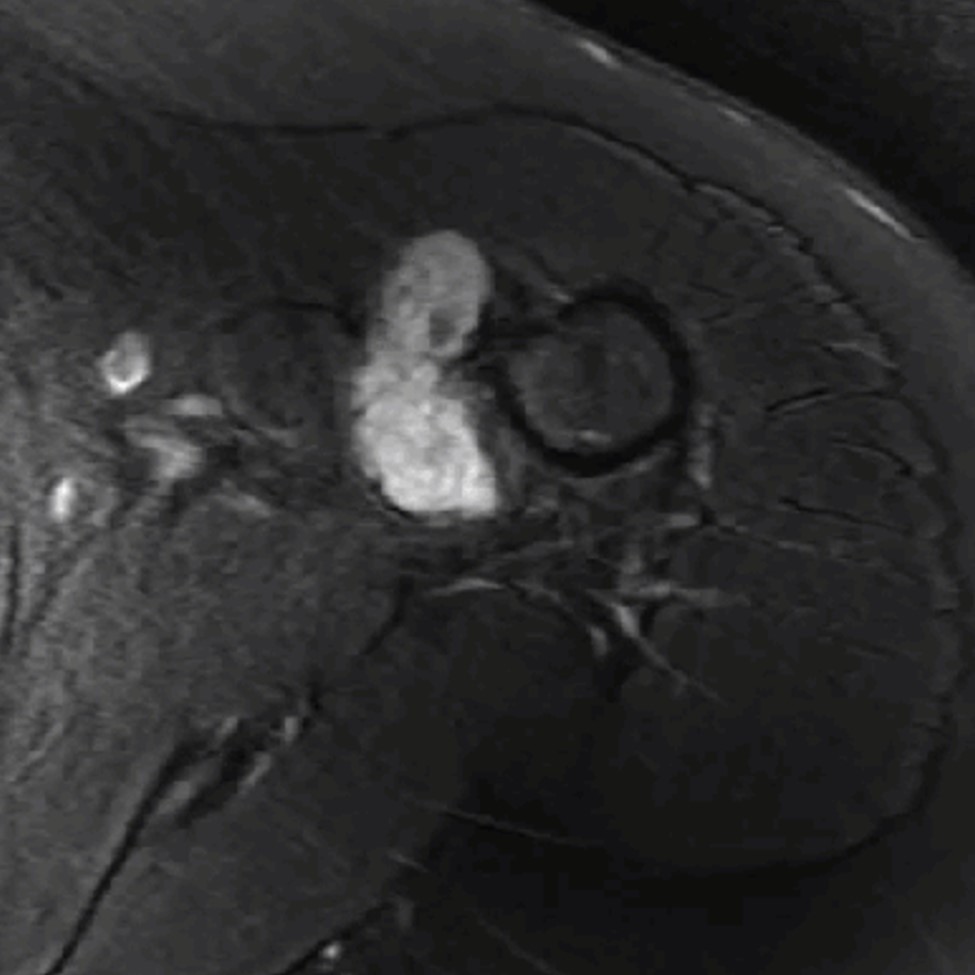
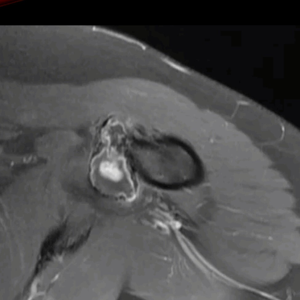
Periosteal chondromas arise from the inner layer of periosteum and can erode the outer table of the cortex. They typically does not extend into the medullary cavity, but they can.
The lesions usually present between the 2nd to 4th decades of life with a 2:1 male predilection.
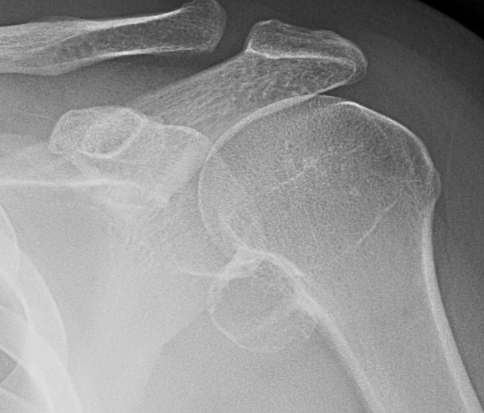
Typical sites include the proximal humerus or distal femur (70%) and phalanges (25%).
11.3.2 Imaging Features
Conventional radiographs show cortical scalloping, rarely associated with intramedullary invasion. A thin periosteal shell with buttress peristeal reaction is typical. Chondroid matrix is often seen.
11.3.3 Differential Considerations
Differential considerations include periosteal osteosarcoma, non-epiphyseal chondroblastoma, and juxtacortical chondrosarcoma.