11.10 Periosteal chondrosarcoma
– Jeremy C. Collette, MD, PhD; Raul Valenzuela, MD
__________________________________________________________________
11.10.1 Background
Periosteal chondrosarcoma, also known as juxtacortical chondrosarcoma, is a malignant neoplasm that is histologically indistinguishable from conventional medullary chondrosarcoma. It is a rare lesions, accounting for 4% of all chondrosarcomas. They are graded into low, intermediate, and high-grade tumors. Most are low to intermediate grade tumors with low rate of metastases (lung is the most common site of metastasis).
They typically occur in the 3rd to 4th decades of life and have a slight male predilection.
The lesions have chondroid matrix and arise from the surface of the bone, lifting the periosteum over themselves as a fibrous pseudocapsule. The underlying cortex is usually thickened or eroded and extension into the medullary cavity is not unusual. Metaplastic ossification is common.
Periosteal chondrosarcomas typically arise from the long bones with a predilection for the posterior aspect of the distal femur.
Treatment is wide excision. Patinet have between 80-90% long-term survival.
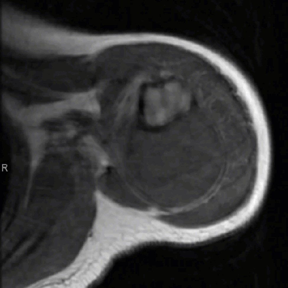
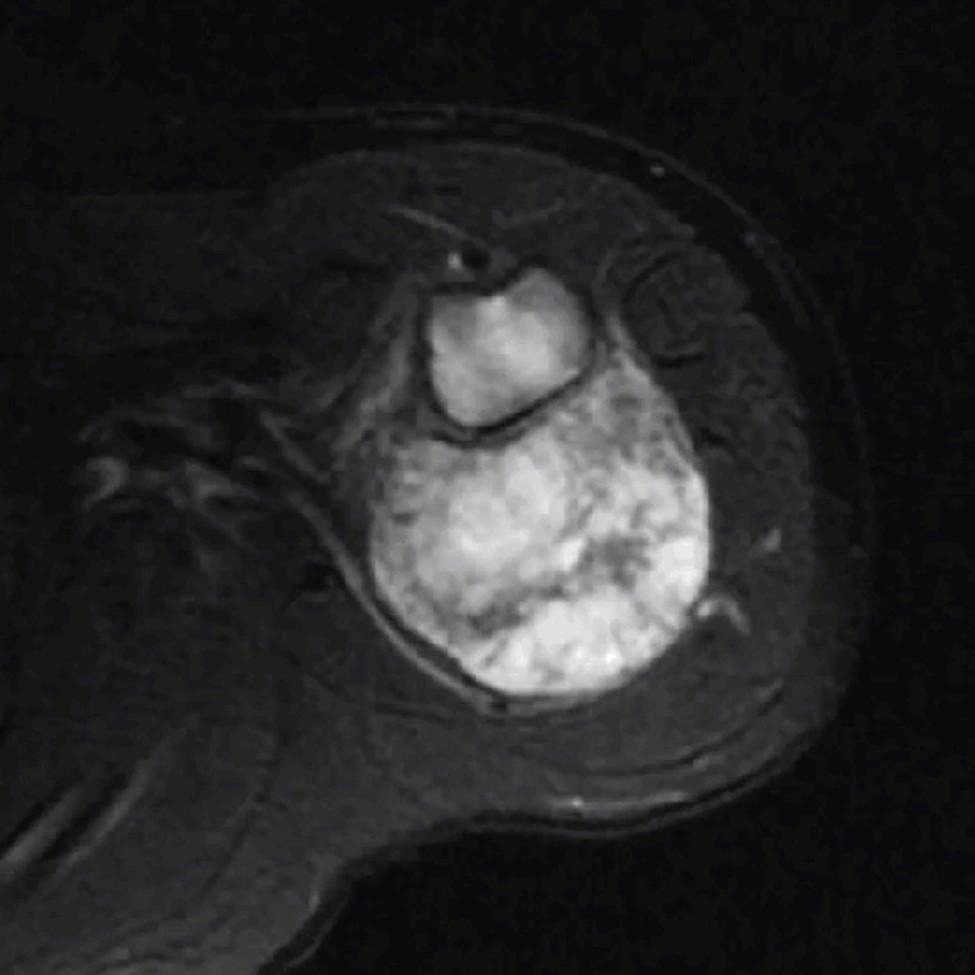
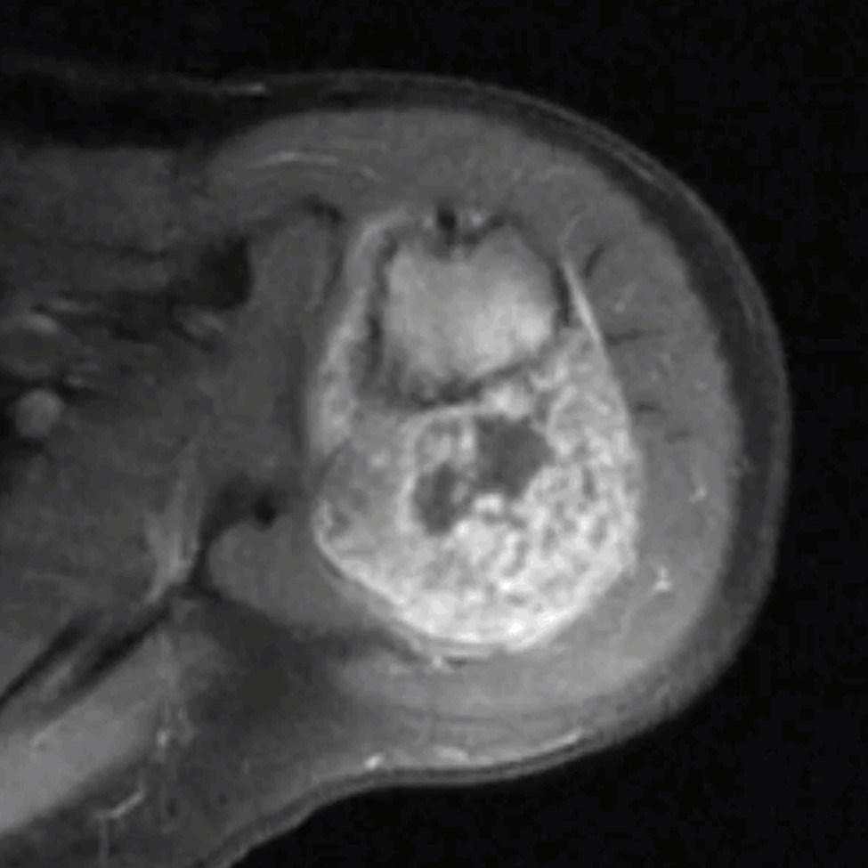
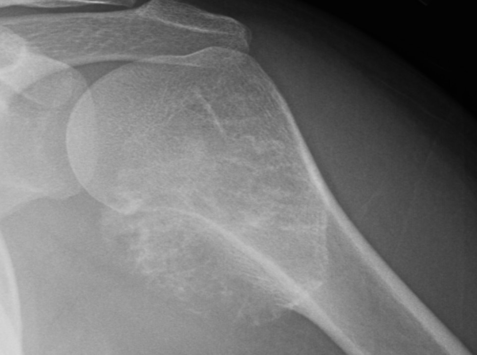
11.10.2 Imaging Features
Radiographs demonstrate a round to oval lobulated soft-tissue mass on the surface of bone (Figure 11.2). Typical chondroid matrix mineralization is usually present. Metaplastic ossification is often apparent to a variable extent. Matrix mineralization is better appreciated on CT. Nonmineralized areas show less attenuation than muscle on CT. On MR, the lesions have low heterogeneous signal intensity on T1-WI, heterogeneous high signal intensity on T2-WI, and peripheral and septal enhancement on T1-WI post-contrast.
11.10.3 Differential Considerations
Differential considerations include, periosteal osteosarcoma, parosteal osteosarcoma, periosteal chondroma, and parosteal lipoma.
Lesion size is the best differentiating feature between periosteal chondroma and chondrosarcomas, with the former being almost invariably smaller (2– 3 cm in size range, 2 cm average) compared to the chondrosarcomas (size range, 3– 14 cm, 5 cm average).
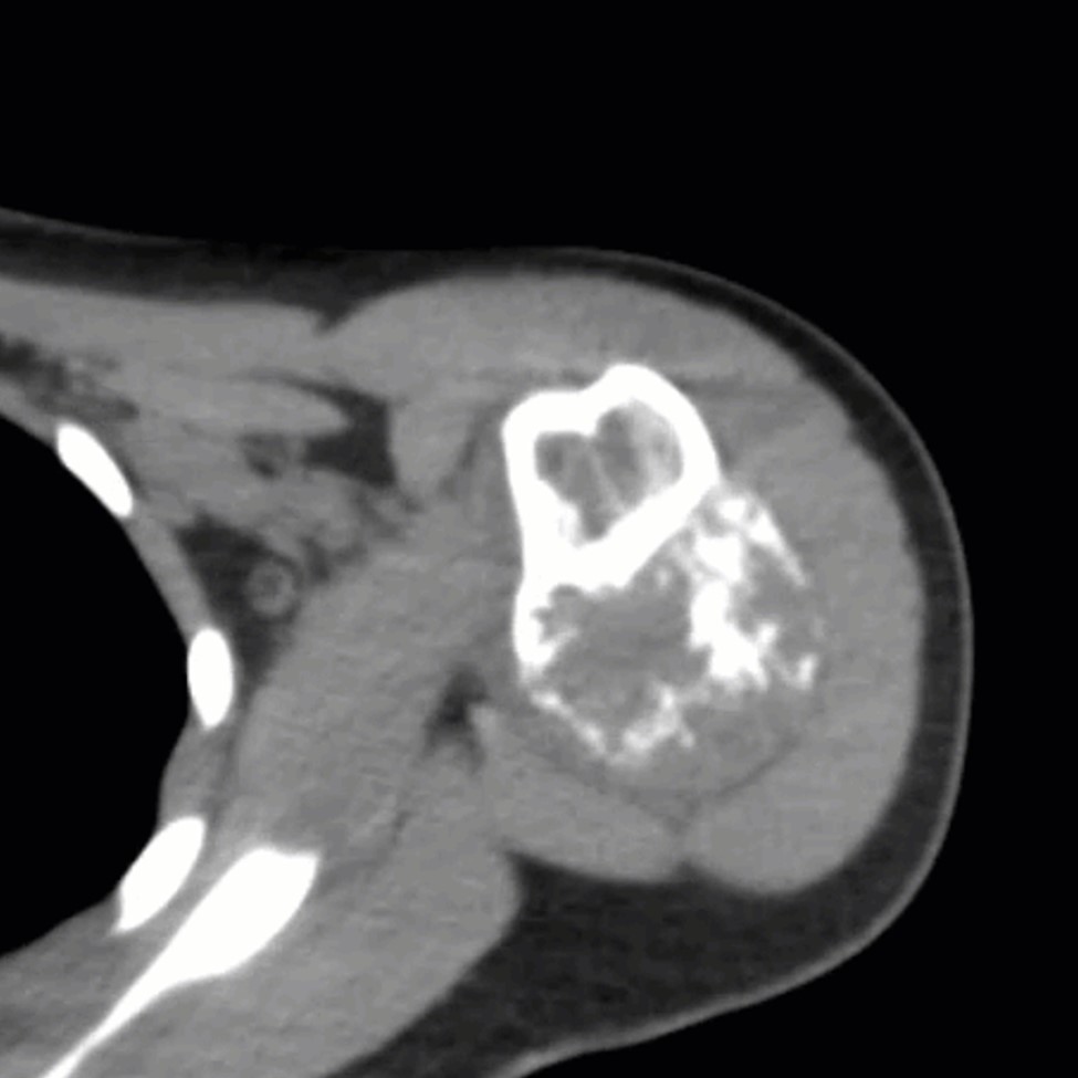 (b) Axial non-contrast CT with soft
tissue windows.
(b) Axial non-contrast CT with soft
tissue windows.
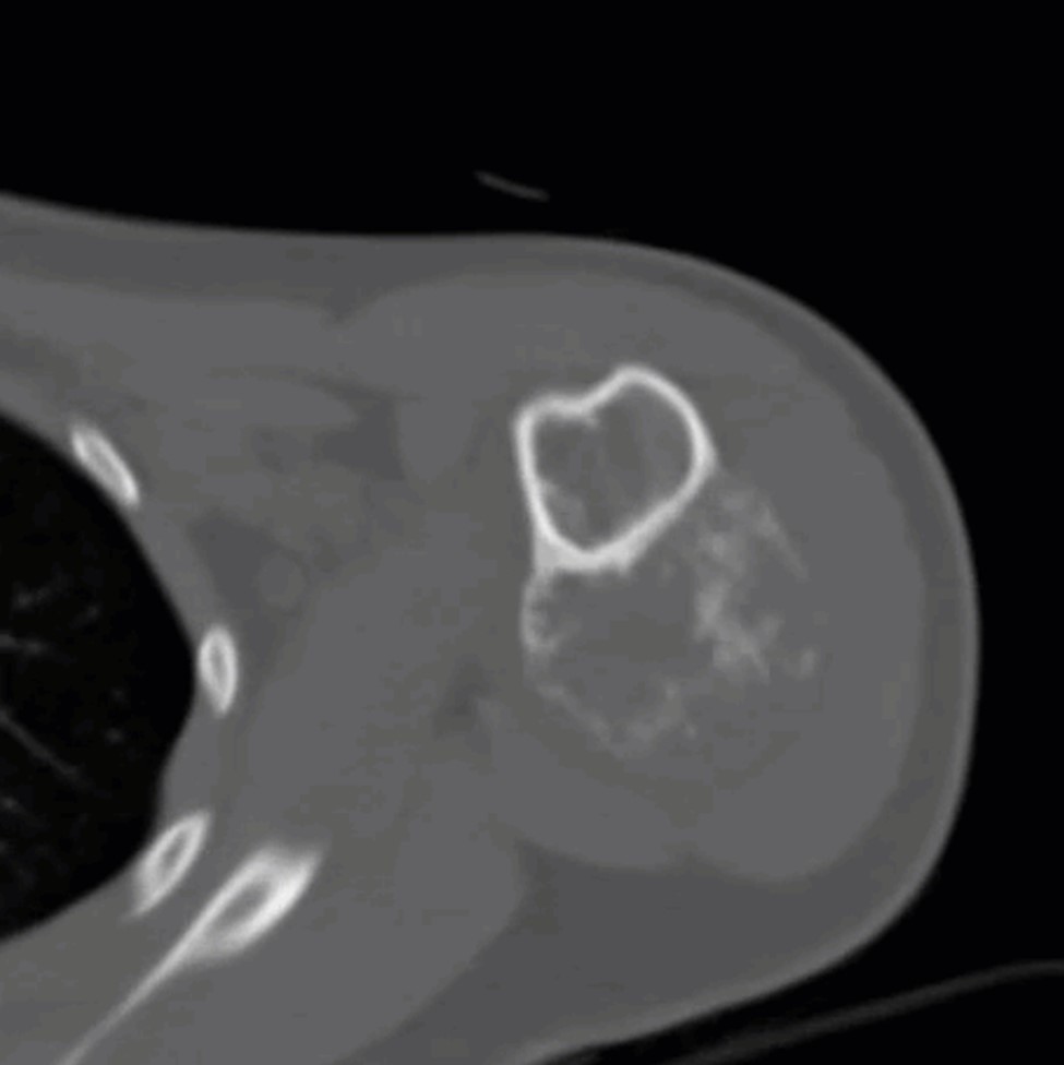 (c) Axial non-contrast CT with sbone
windows
(c) Axial non-contrast CT with sbone
windows